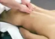
Sciatic nerve neuralgia is considered to be one of the most difficult abnormalities of the support and movement system. Clinical expressions of sciatic nerve neuralgia include sharp pain irradiating to the lower extremities which increases during any of the patient's attempts to move and of course limited range of motion on all axes and planes. Usually, when a patient appears at the doctor's office with such a difficult clinical picture, in addition to prescribing painkillers, anti-inflammatory drugs, and/or utilizing injections, the doctor will usually refer the patient for radiological examination. Today, the most common radiological examinations are the MRI and/or CT scan. In many cases, MRI test detects bulging and/or herniation of discs. If symptoms are not alleviated in a period of a few weeks due to conservative methods of treatments (above mentioned oral or injected medication) consideration will be given to surgical intervention. The problem is that in many cases, this difficult neurological picture is not the result of the disc herniation, but is actually the result of piriformis muscle syndrome.
Anatomy of the Piriformis Muscles
The Piriformis muscle originates from the anterior surface of the sacrum and inserts into the greater trochanter. It shares the passage through the greater sciatic foramen wit several important nervous and vascular structures including nerves which provide innervation to pelvic inner organs, the gluteals, and the lower extremities The piriformis muscles can accumulate tension to the point that they-will start compressing/ impinging sciatic nerve and awaking in some cases intolerable pain in the buttocks and lower back with irradiation to the extremities.
Sciatica nerve neuralgia can be the result of compression of the S1 spinal nerve as well as due to the compression of the sciatic nerve by over-tensed piriformis muscles. If patients with symptoms of sciatica have the ability to bend forward without awaking pain, most likely the sciatica neuralgia is the results of piriformis muscle syndrome.
The Causes for Accumulation of Tension in the Piriformis Muscles
- Due to spondylosis, including herniation of disc, spinaI nerves can be irritated (please do not confuse irritation of the spinal nerve with compression of the spinal nerve.) Irritation of the spinal nerves that provide innervation to the piriformis muscles (S1 spinal nerve) will cause a gradual accumulation of tension in these muscles, to the point that they will compress the sciatic nerve.
- Physical overload of the muscles such as vigorous exercise, job performances that put too much static load on the piriformis muscles, hormone changes, exposure to toxins, physical and/or psychological trauma, etc.
Once, I presented to a group of doctors on medical massage and I mentioned the importance of medical massage treatment in cases of sciatic nerve neuralgia. I also pointed out that in many cases, herniation of the intervertebral disc does not play the main cause for this difficult pathology. One of the young doctors who attended my presentation said to me "According to you, many spinal surgeries are performed unnecessarily," to which I replied, "This is true." This young doctor asked me the following question: "How can I explain that after surgery this terrible irradiating pain to the extremities disappeared?" I replied, "Under total anesthesia, all muscles drop any tension they were holding. Thus, if the cause of this neurological picture was over-tensed piriformis muscles, the post-anesthesia patient experiences immediate relief from the shooting pain to the lower extremities. Massage therapy procedure in cases of sciatic neuralgia is directed at reducing the tension in piriformis muscles, which in turn will impinge/compress nerve less, as well as to eliminate trigger points that possibly can be developed on the pathways of sciatica nerve and branches. The protocol of massage therapy that I offer you was initially developed and proposed by professor Popelansky, MD. As I mentioned before, spondylosis, including degenerative disc disease, herniation of disc, spondyloarthritis, etc, can irritate the spinal nerves and cause reflexively increased tension in piriforms muscles and others. Therefore, it is very important to start treatment of sciatica neuralgia from the lumbar spine area, sacra-iliac crest region. The proposed segment-reflex massage will not only cause reduction of tension in the piriformis muscles before we even begin working on them, but also will increase arterial blood supply to the region. It is very important to place a pillow under the stomach of the client (as he/she lays face down). Pillow should be located on the projection and level of lumbar spine to prevent hyperextension and possible protective contraction phenomenon of the muscles at the time of applying moderate pressure on the lumbar spine region.
Steps for Piriformis Muscle Syndrome (Sciatica Neuralgia) Relief
Pathogenesis:
The piriformis muscle has an unusual anatomy. It originates from the anterior surface of the sacrum in the area of the sacro-iliac joint, exits the pelvis via the greater sciatic foramen, and iserts into the greater trochanter. The greater sciatic foramen is formed by the sacrum, the iliom bone and the sacrospinous ligament. Important vascular and nervous structures are located along the piriformis muscle. When the piriformis muscle leaves the pelvis, it divides the greater sciatic foramen on the suprapirifomis and infrapiriformis recesses. The gluteal superior nerve and artery exit the pelvis via the suprapiriformis recess. The gluteal inferior nerve and artery, the pudendal nerve and artery, and the sciatic nerve exit the pelvis via the infrapiriformis recess. Besides that, the sacral plexus is located in the pelvis on the anterior surface of the piriformis muscle. This plexus is the origin of many nerves (i.e. sciatic, gluteal inferior, cutaneous posterior) which support the innervation of pelvic inner organs, the gluteal area, and the lower limb. The practitioner has to remember that often the sciatic nerve goes through the piriformis muscle. This fact can make the clinical picture and the prognosis of treatment of PMS worse. After exiting the pelvis, the nerves whish originated in the sacral plexus run between the piriformis muscle and fascia which cover the gluteus maximus muscle. This is why the increasing tones of the piriformis muscle evokes a rich clinical picture in the gluteal area and in the lower limbs. Actions of the piriformis muscle are abduction and lateral rotation of the thigh. The piriformis muscle receives motor innervation from the S1-S2 spinal nerves. Thus, chronic irritation or compression of the S1-S2 spinal nerves can cause PMS. Static or dynamic overload of the piriformis muscle evokes the same clinical picture.
Clinical Symptoms:
A special sign of PMS is a combination of lumbosacral pain, local pain in the piriformis muscle and different compression syndromes. Patients complain about a dull, pulling pain in the lower back, gluteal area, regions of the sacro-iliac, and hip joints. Pain increases during standing, squatting, or when crossing one's legs. Pain radiates to the groin, hip joint, and posterior surface of the knee joint. The patient also shows clinical signs of irritation or direct compression of the nervous and vascular structures which are located nearby and are affected by the overtensed piriformis muscle. The examiner can see vasomotor reactions (vasospasm or vasodilatation) as well as symptoms of the peripheral nerves involvement.
Compression of the S1 spinal nerve by the intervertebral disk and irritation of the sciatic nerve due to an overtensed piriformis muscle are two major causes of sciatica. It is very important to make the correct differentiation between these two causes and to precisely target medical massage treatment. In the case of lumbago or lumbalgia, pain and restriction of movements in the lumbar spine are the main symptoms. In this case, pain is more intense, radiating all the way down to the plantar surface of the foot and toes. A diagnostic examination shows a muscular spasm in the lumbar part of the back as well as in the gluteal area. In the case of piriformis muscle syndrome, the sciatica pain is dull and patients are more mobile. Their range of movement in the lumbar part of the spine is unchanged or only slightly restricted. The pain is located in the gluteaI area, radiating down to the leg, but most likely to the level of the lateral surface of the lower 1/3 of the leg. During the diagnostic examination, the examiner will not find any significant pain or spasm in the lumbar part of the back. Local tension and severe pain can be found during palpation in the gluteal region especially in the areas of the trigger points.
- Place both thumbs on either side of the spine at the L/S region. Under pressure, (freccion action), from L/S region - T12/L5 region, massage the paravertebral zones 8-10 times. (Optional - massage with thumbs in circular motion)
- Standing in front of the lower back, perform petrissage #1 and #2 on all areas from L1 to S5. Petrissage must be performed on the glutes muslces, as well as transmitted pressure to Piriformis muscles. All areas must be checked for trigger points. With fingers bent at 90 degrees, use fingertips to palpate all areas of the lower back to detect trigger points; pay attention to areas of the iliac crest, the paravertebral zones and the quadratus lumborum. All discovered trigger points must be ischemically compressed for 30 seconds each.
-
Automatically, we can assume that in the area of a trigger point, arterial blood supply is significantly decreased. By gradually compressing the trigger point and reaching the threshold of pain, we additionally decrease this blood supply to the point. After 10 seconds of this ischemic compression, we ask our client to report their ability to tolerate additional compression (please do not overdo this pressure; stop at a pressure level that your client indicates is sufficient, and do not increase it. Do not increase the pressure if muscles demonstrate protective contraction to the pain sensation.) After 20 additional seconds of compression, quickly withdraw your fingers. While performing ischemic compression of trigger points, arterial blood will accumulate around the compressed point. With the fast withdrawal, arterial blood will rush to the area of the trigger point, and a vasodilation reflex will be awakened.
- Perform petrissage #4 on paravertebral zones starting from lumbar-sacral region and
finishing with L1-T12 level. Description of petrissage #4. With the left or right hand extend the thumb so that it will form a 90 degree angle with the rest of the fingers and place the hand on the paravertebral zone. In circular motion, with the upper parts of the second, third and forth fingers on the left hand shift tissue toward the inside part of the formed 90 degrees angle on the right hand. Hands are interchangeable.
- With the backs of the fingers, towards the heart only, massage the paravertebral zones in region of the lower back. (Please do not massage the spinous processes).
- Palpate with the tips of the fingers, the L5 spinous process. From this point, move finger laterally (away from spine) to the soft tissue area next to it. Replace finger with elbow. Applying initial pressure, perform long strokes under freccion action around the pelvis, removing the glutes, and stimulating Piriformis muscles. Repeat this motion 8 times.
- Perform petrissage number 5 on all areas of the lumbo-sacral region, including the paravertebral zones, quadratus lumborum, glutes, and Piriformis.
- Options: leg straight, OR, leg bent the knee, while thigh is abducted and laterally rotated at the hip joint. Place the elbow in the area of the greater trochanter. Shift the glutes medially, and apply massage under angled pressure to stimulate the piriformis muscle. Discovered trigger points may be ischemically compressed with the elbow.
- All pathways of pain must be checked for possible development of trigger points. Ischemic compression of trigger points on the posterior thigh can be done with the point of the elbow (when arm is flexed).


